Monday, January 31, 2005

ILC with extensive LCIS
ILC with extensive LCIS ILC with extensive LCIS

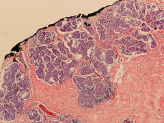
These pictures were taken from the margins of a breast conserving specimen that was done because of invasive lobular carcinoma (ILC) (diagnosis was established by punch biopsy). Only lobular carcinoma in situ (LCIS) is shown here. The diagnosis after breast conserving surgery was ILC with extensive LCIS. Excision was incomplete regarding the LCIS, therefore re-excision was recommended (G-292/05).

Comment: As you can see on the photomicrographs, the LCIS fills the entire lobular system and is also present in the margins. While it is commonly argued, that LCIS in the margins is no justification for re-excision, this is not the case when the LCIS is very extensive. Under these circumstances it is likely that you find more extensive LCIS in the remainder of the breast and possibly multifocal invasive foci. Therefore, a re-excision may be warranted when you find extensive LCIS.
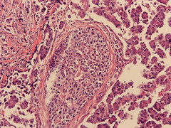
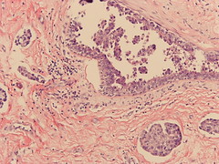
Invasive micropapillary carcinoma of the breast

more on micropapillary breast ca. more on micropapillary breast ca.

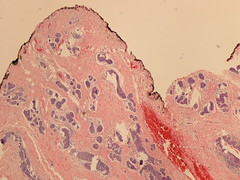
On the left you see an invasive carcinoma of the breast with micropapillary differentiation and intraductal component in the center at medium power view (10x). This picture was taken from a breast conserving specimen of a 3 cm carcinoma with extensive lymphovascular invasion and lymph node metastasis. The intraductal component and the lymphangic spread are seen on the right.

Micropapillary carcinoma are notorious for extensive lymphovascular spread (Pettinato G et al. Am J Clin Pathol. 121: 857-66, 2004).

Mucinous adenocarcinoma (macro)

Mucinous adenocarcinoma (macro)

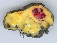
Just a nice macro image of an invasive mucinous adenocarcinoma of the breast. Unfortunately, there is no ruler in this picture, but the size of the tumor is about 1 cm (G-00337/05). Note: This picture was taken with a flatbed scanner just by placing the sliced specimen on the glas and closeing the lid (Agfa Arcus 1200 scanner).

Sunday, January 30, 2005

Erosive papillomatosis of the nipple

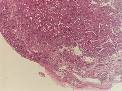
This is a nice case of erosive papillomatosis of the nipple. The papillary structures fill almost the entire picture, and you can see the epithial erosion at the left hand side. Unfortunately I have no images of immunohistology yet.

The image was taken with a Nikon DN100 camera on a Nikon Optiphot 2 Microscope at 4x.

What is this blog about

This blog is intended as a personal notebook to record rare and memorable cases in gynaecopathology. Not infrequently, I would like to go back to a certain case quickly, but it may be difficult to retrieve it. This is an attempt to store cases prospectively as a future reference.

Comments are welcome, of course. But no questions from people who are not familiar with these entities, please.