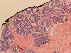
ILC with extensive LCIS
|
|
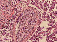
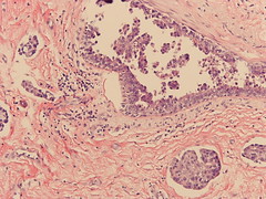
These pictures were taken from the margins of a breast conserving specimen that was done because of invasive lobular carcinoma (ILC) (diagnosis was established by punch biopsy). Only lobular carcinoma in situ (LCIS) is shown here. The diagnosis after breast conserving surgery was ILC with extensive LCIS. Excision was incomplete regarding the LCIS, therefore re-excision was recommended (G-292/05).
Comment: As you can see on the photomicrographs, the LCIS fills the entire lobular system and is also present in the margins. While it is commonly argued, that LCIS in the margins is no justification for re-excision, this is not the case when the LCIS is very extensive. Under these circumstances it is likely that you find more extensive LCIS in the remainder of the breast and possibly multifocal invasive foci. Therefore, a re-excision may be warranted when you find extensive LCIS.
posted by P.S. at 1/31/2005 06:26:00 PM
![]()